TLDR;
This video provides a comprehensive overview of the skeletal bones, starting from the skull and moving down to the toes. It covers the cranial and facial bones, vertebrae, thorax, upper limb, pelvis, and lower limb, including the ankle and foot. Key points include the specific bones in each region, their connections, and unique features like the true and false ribs, the structure of the hand and foot, and the joints between bones.
- Cranial bones protect the brain, while facial bones form the structure of the face.
- The vertebral column consists of cervical, thoracic, and lumbar vertebrae, as well as the sacrum and coccyx.
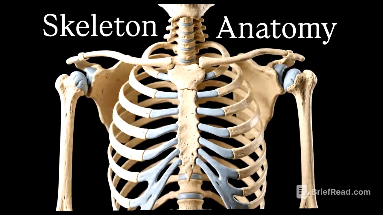
- The thorax includes the clavicle, scapula, sternum, and ribs, with true ribs connected directly to the sternum via coastal cartilage and false ribs not directly connected.
- The upper limb comprises the humerus, radius, ulna, carpals, metacarpals, and phalanges, forming joints like the elbow and shoulder.
- The pelvis is made up of the ilium, ischium, pubis, and sacrum, connecting to the femur at the acetabulum.
- The lower limb includes the femur, tibia, fibula, patella, tarsals, metatarsals, and phalanges, forming the knee and ankle joints.
Introduction to Skeletal Bones [0:00]
The video introduces a discussion on skeletal bones, covering the entire body from the skull to the toes. The presenter, Rashil, starts with the bones of the head, beginning with the cranial bones that protect the brain.
Cranial and Facial Bones [0:15]
The frontal bone is highlighted as a key cranial bone. Connected to it is the sphenoid bone, located inside the skull and referred to as the butterfly bone. The ethmoid bone, a sponge-like structure, sits between the facial bones. The parietal bone connects to the frontal bone, and the occipital bone forms the base of the skull. Moving to the facial bones, the nasal bone is identified as a delicate structure prone to fracture. The maxilla and zygomatic bones (cheekbones) are also pointed out. The mandible bone connects to the temporal bone via the temporomandibular joint (TMJ).
Vertebrae [1:26]
The video explains the vertebral column, which includes seven cervical vertebrae (C1-C7), twelve thoracic vertebrae (T1-T12), and five lumbar vertebrae. The sacrum and coccyx are also mentioned. The first cervical vertebra (C1) is called the atlas, and the second (C2) is called the axis.
Thorax [2:21]
The thorax section begins with the clavicle, a long bone connected to the scapula and sternum. The clavicle joins the manubrium at the sternoclavicular joint. The sternum consists of the manubrium and sternum body, with the joint between them known as the sternal angle. There are 12 pairs of ribs, connected to the sternum via coastal cartilage and to the vertebrae in the back. The first 10 ribs are true ribs, directly connected to the coastal cartilage, while the 11th and 12th ribs are false or floating ribs, not directly connected. The xiphoid process is a small bone connected to the sternum.
Upper Limb [3:39]
The upper limb section revisits the clavicle and introduces the scapula. The shoulder joint is formed by three bones: the humerus, scapula, and clavicle. The humerus is a long bone that connects with the radius and ulna at the elbow joint. The elbow joint is thus made up of the radius, humerus, and ulna. The head of the humerus connects to the glenoid fossa of the scapula, forming the glenohumeral joint.
Hand Bones [5:07]
A closer look at the hand bones reveals the ulna and radius. There are eight carpal bones in the wrist, followed by five metacarpals. The phalanges consist of proximal, middle, and distal parts, except for the thumb, which only has distal and proximal phalanges. The joints between the fingers include the metacarpophalangeal (MCP) joint, proximal interphalangeal joint, and distal interphalangeal joint.
Pelvis [6:30]
The pelvis is composed of the ilium, ischium, pubis, and sacrum. The ilium is shown in red, the ischium in yellow, and the pubis in green. The two pubic bones are joined by the pubic symphysis. The sacrum and ilium connect at the sacroiliac joint. The acetabulum is the socket where the head of the femur joins the pelvis.
Lower Limb [7:24]
The lower limb section starts with the femur, the longest and toughest bone in the body, which joins the pelvis at the acetabulum. The tibia is in the medial position, and the fibula is in the lateral position. Medial means towards the body's center, while lateral means away from the center. The patella, or kneecap, covers the knee.
Ankle and Foot [8:12]
The ankle and foot contain seven tarsal bones: the talus, cuboid bone, navicular bone, three cuneiform bones, and the calcaneus bone. There are five metatarsal bones and phalanges, similar to the hand. The phalanges have proximal, middle, and distal parts, except for the thumb, which only has distal and proximal parts.